Radiography is a vital part of dentistry. Without it, dentists couldn’t see what’s going on deep inside the mouth during an exam. In an oral care practice, it’s a dental assistant’s responsibility to prepare patients and take X-rays as part of an examination.
What is an X-ray?
X-rays are used in the medical and dental fields to capture images to diagnose health concerns. They utilize an element called tungsten which, when bombarded with electrons, produces high-energy electromagnetic waves or X-rays, that pass through soft tissue, like skin, but are absorbed by dense tissue, like bone. Discovered accidentally more than a century ago by German scientist Wilhelm Roentgen, radiography has been a mainstay in medicine ever since.
Why are X-rays Done?
X-rays, or radiographs, are performed during an exam to allow dentists to see teeth, nerves and bone below the gum line. They’re used both preventively and to diagnose dental issues when patients have symptoms such as pain or tooth sensitivity.
X-rays can detect a broad range of dental issues, including:
- Cavities
- Abscesses
- Loose crowns and fillings
- Changes in the root canal
- Impacted wisdom teeth
- Irregular bites
- Periodontal bone loss
- Abnormal tooth development
- Cysts
- Tumors
- Jaw disorders such as arthritis or Temporomandibular Joint Dysfunction (TMJ)
Radiographs also help dentists plan restorative care, such as dental implants.
Types of Dental X-rays
There are five common types of X-rays done in dental offices, each having a specific use or exam purpose. They include periapical X-rays, bitewing X-rays, full mouth X-rays, occlusal X-rays and panoramic X-rays.
Periapical X-rays
Periapical X-rays capture an image of the whole tooth from the crown above the gumline to the root end. Used to highlight only one or two teeth at a time, they provide a top to bottom view that helps diagnose painful conditions or changes in teeth resulting from periodontic treatments.
Taking a periapical X-ray requires placing a small strip of film in a plastic holder into the mouth. Attached to the holder is a frame with a ring that mirrors the position of the film, helping the dental assistant align the X-ray machine opposite the tooth. Patients bite down on the holder to keep the film steady while taking the X-ray.
Bitewing X-rays
Bitewings are similar to periapical X-rays, but they capture full sections of both the upper and lower molars and bicuspids. They’re used both diagnostically and preventively, detecting issues from tooth decay to periodontal disease, and also showing how the top and bottom teeth occlude.
The process is similar to taking periapical X-rays, but the film is larger. Newer digital systems use smaller X-ray sensors instead of conventional film. The results are the same, but the procedure is more comfortable for the patient.
Full Mouth X-rays
Full mouth X-rays include both periapical and bitewing radiographs. They give the dentist a baseline impression of a patient’s oral health that can be used later for comparison when problems occur.
Occlusal X-rays
Occlusal X-rays show the roof and floor of the mouth. They’re used primarily by pediatric dentists to help monitor the development of teeth that haven’t yet broken through the gums or to check for congenital conditions and injuries. In adults, a dentist may order them to help find tumors or cysts. The process is similar to a bitewing.
Panoramic X-rays
Unlike other types of dental radiographs, panoramic X-rays are extraoral, no film goes into the mouth. Instead, a rotating arm captures a two-dimensional image of the entire mouth from ear to ear.
Panoramic X-rays help diagnose, to assess bite, as well as dental issues that may extend to the jawbone. They’re regularly used by orthodontists and dental surgeons to plan implants, dentures, braces and extractions. The procedure requires no special preparation.
Patients typically have full mouth or panoramic X-rays done during their first examination and then again, every 3–5 years. Since posterior teeth are more likely to have invisible decay, bitewings are done preventively every few years based on the patient’s risk. More specific radiographs are taken as needed to diagnose the cause or dental symptoms.
For certain conditions, specialty X-rays may be necessary. Large dental practices may have the equipment to do them, but smaller practices may have to refer patients to a hospital or imaging center.
Specialty Dental X-rays
There are a few X-rays used in special situations, when additional examination of the teeth is needed. They include Cephalometric projections, Sialography, and computerized tomography.
Cephalometric Projections
This type of X-ray shows the entire side of the head in proportion to the patient’s profile. They help orthodontists plan treatments that achieve a natural look.
Sialography
Dentists use sialography to diagnose problems with salivary glands. A dye is injected into the organs so that they’re visible on X-rays. The dentist typically does the injection, but a dental assistant may take the film.
Computerized Tomography
X-rays produce two-dimensional images; CT scans offer a three-dimensional view of oral structures. They’re particularly useful for planning the size and location of dental implants.
The Dental Assistant’s Role
In private practices, dental assistants are responsible for preparing patients for their examination and X-rays. Getting the highest-quality images requires understanding how X-ray equipment works and how body positioning affects results. Dental assistants are trained to follow safety protocols and to ensure patients are fully informed before examination and testing.
Preparation for X-rays includes equipment checks, verifying orders, patient education screening, physical preparation and reviewing results.
Equipment Checks
Before the patient arrives, dental assistants should check equipment to ensure it’s functioning correctly, and film should be preloaded into holders to save time. Any devices that come into contact with a patient’s body fluid must be sanitized before and between appointments.
Verifying Orders
To minimize exposure to radiation, dentists only order the X-rays needed for optimal oral care. It’s up to the dental assistant to check orders and take only the necessary radiographs.
Patient Education
Since X-rays emit small amounts of ionizing radiation that can damage DNA, patients often ask if they’re safe. Generally speaking, they are– especially with the advent of digital X-ray machines that produce less radiation. However, minor repeated exposures can add up over time, and patients have the right to know how this could affect their health. The dose of radiation for a full set of dental X-rays is about the same as one would be exposed to during a short flight.
Dental assistants can ease patients’ minds by explaining the risks, and the protective measures used to minimize exposure to radiation. Demonstrating how protective lead-lined aprons help to shield the body during X-rays is reassuring.
Patients will also want to know what taking X-rays entails. They’re not painful, but they do require cooperation. Patients are asked to bite on film holders and hold their breath while radiographs are taken, in order to decrease artifact distortions such as blurring or dark spots that affect the clarity of the image. Well-informed patients who know what to expect are usually cooperative and this is helpful for creating clear, useful x-ray images.
Screening
Before taking X-rays, dental assistants screen patients for higher than average health risks from radiation. Risks are extremely low for the general population, but they’re slightly higher for pregnant women and patients under 30 years-old. Studies correlate radiation with an increase in thyroid cancer in children and young adults, so a lead-lined collar is used to protect patients against thyroid injury.
Taking X-rays also requires placing film in the mouth, which is something that can be uncomfortable for a patient with a toothache. As part of an examination, dental assistants screen for symptoms so they can alter its position whenever possible to limit discomfort.
Physical Preparation
Most X-rays are taken while the patient sits in a dental chair awaiting examination, while panoramic X-rays may require the patient to stand. Metal objects, including jewelry and glasses, could interfere with the final image so these are typically removed when preparing the patient for the X-ray. Patients should be reminded to stay still for the few seconds it takes to develop the image, to increase clarity.
Once the equipment is ready, patients are draped with lead aprons and neck collars, and the X-ray machine is moved into place. From start to finish, limited X-rays take only a few seconds to complete. Including preparation, panoramic x-rays may take up to five minutes.
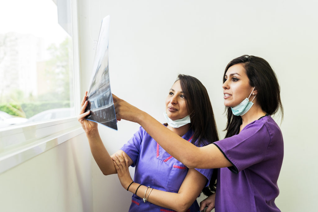
Reviewing Results
The dentist’s time is valuable, so dental assistants check X-rays before presenting them to the dentist for examination. Distorted images should be retaken.
Final Thoughts
Radiography revolutionized dentistry, and it remains the most important diagnostic tool for dentists today. It’s an integral part of a comprehensive examination and one of the many ways dental assistants help provide top-quality oral care.
Did learning about taking X-rays and examination interest you? Do you need to become a dental assistant first? The Dental Assistant training program at Meridian College provides extensive hands-on training including a school externship at a dental office where you will assist the dentist in treating actual patients.
Contact Meridian College today to learn more about becoming a dental assistant.