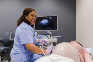
 Healthcare is a team effort. Doctors collaborate with other licensed clinicians and allied health professionals to bring the broadest expertise on the patient’s behalf. Ultrasound technicians play an indispensable role in this process by helping physicians see what they otherwise couldn’t, such as detailed images of internal body structures. Also known as diagnostic medical sonography, it’s a critical science and a rewarding career.
What Does an Ultrasound Technician Do?
Ultrasound is a non-invasive diagnostic imaging technique that uses sound waves to see organs, blood vessels, and soft tissue. Ultrasound technicians manage the entire sonography process, from patient engagement to documenting test results.
Their primary responsibilities include:
Patient Preparation
Preparation for an ultrasound varies based on the images ordered. Still, patients should understand the test’s purpose and what to expect, including how long it will take and if discomfort is likely.
Depending on the type of sonogram, patients may need to lie down, sit up, or assume a side-lying position to provide access to the affected area. Reviewing the procedure step-by-step will ensure the patient’s cooperation with your instructions.
Making the patient as comfortable as possible using pillows or support devices is a must; some exams take up to 30 minutes. You’ll communicate with the patient throughout the procedure, answering questions and providing reassurance.
Conducting Sonograms
Sonographers operate ultrasound equipment, including the transducer, a handheld device that produces high-frequency sound waves, and the monitor that captures the images.
The process begins by applying a water-based gel to the patient’s skin to facilitate the transmission of sound waves. Filling in air gaps reduces image distortions while protecting the patient’s skin against friction as the transducer is moved to optimize images.
As sound waves penetrate the body, they bounce back as they encounter different body structures, creating an echo that generates images on a monitor. Ultrasound technicians adjust machine settings in real-time, manipulating the depth and frequency of sound waves to obtain the most comprehensive pictures.
Image Analysis
Radiologists review ultrasound images for quality, clarity, and completeness, creating reports that identify and describe the condition of body structures. Using measurement tools in the ultrasound software, they can note organ dimensions, flow velocity through blood vessels, and many more parameters.
Ultrasound technicians, however, must be adept at interpreting images during an ultrasound exam or risk missing abnormalities. Both the radiologist and the referring physician rely on their expertise.
Clean Up
Ultrasound technicians assist patients with post-exam cleanup, wiping gel from their skin and providing supplies for cleansing sensitive areas. Between patients, you’ll maintain the changing areas, restock your workstation, and sanitize equipment to prevent the spread of infection.
Patient Education
Patients are naturally curious about their test results, so ultrasound technicians can provide general information about the findings without being too detailed. Educating patients about the next step in the diagnostic process helps ease their anxiety.
How Do You Become an Ultrasound Technician?
Becoming a medical sonographer requires a higher education. Students can choose from degree or diploma programs, but most choose vocational training for its accessibility and convenience. Students graduate work-ready and prepare for certification in less time than their college-educated peers. It’s a fast track to a new and rewarding career without spending four years in a classroom.
What Do You Learn During an Ultrasound Technician Program?
Sonography is a straightforward yet complex procedure that requires technical expertise, a firm grasp of ultrasound science, sound clinical judgment, and people skills, all things you’ll learn in a vocational school sonography program.
The curriculum covers:
Anatomy and Physiology
Anatomy and physiology courses for ultrasound technicians explore the structure and function of the human body as it relates to sonography.
Topics include:
Medical terminology — the medical terms used to describe anatomy, physiology and pathology related to ultrasound examinations.
Body structure — how the body is made from the cellular to the systemic level, including major structures and organ systems.
Physiology — basic biochemical processes and how organs work together to support life.
Pathophysiology — abnormal physiological processes affecting the structure and function of organs and blood vessels.
Principles and Protocols of Sonography
This course expands on basic anatomy and physiology, delving into the specifics of ultrasound technology, the principles of imaging and protocols for different types of sonograms.
Students learn about:
Ultrasound anatomy — how structures appear in various imaging modalities, including ultrasound.
The ultrasound technician’s role — a review of the ultrasound technician’s responsibilities and professional expectations.
Scanning protocols — methods for optimizing image quality for abdominal, gynecological, obstetric, vascular, and cardiac sonograms.
Patient positioning — positioning techniques based on the type of ultrasound and the patient’s body morphology.
Acoustical Physics and Instrumentation
Students learn the fundamental principles and concepts related to ultrasound equipment and imaging.
Concepts include:
Basics acoustics — the fundamentals of sound waves, including wave properties, frequency, wavelength, amplitude, and velocity.
The Pulse-Echo Principle — how echoes are processed to generate visual representations of internal structures.
Image formation — the principles of image formation, including beam steering, focusing and the creation of cross-sectional images.
The Doppler Effect — an introduction to Doppler technology and the interpretation of Doppler waveforms.
Artifacts — preventing unwanted distortions in ultrasound images, such as shadowing, reverberation, and acoustic enhancement.
Ultrasound instrumentation — the components of ultrasound equipment, including the control panel, monitor, and transducer.
Cross-Sectional Anatomy
Ultrasound requires the interpretation of cross-sectional images. This course helps students see the body from a whole new perspective.
You’ll study:
Imaging modalities — the imaging methods that produce cross-sectional images, including ultrasound, CT, and MRI, including their advantages and limitations.
Directional and positional terminology — the terms used to describe the location of structures on different anatomical planes.
Thoracic anatomy — a detailed look at thoracic anatomy and associated structures, including the lungs, heart, mediastinum, and major blood vessels.
Abdomino-pelvic anatomy — an in-depth study of the cross-sectional abdominal and pelvic anatomy, including the liver, spleen, kidneys, gastrointestinal organs, and reproductive organs.
Musculoskeletal anatomy — an examination of the cross-sectional anatomy of the musculoskeletal system, including bones, joints, muscles, and soft tissues.
Cross-sectional blood vessel anatomy — the anatomy of major blood vessels, including arteries and veins.
Abdominal Sonography
Ultrasound is a common imaging modality for a broad range of abdominal disorders.
This course covers:
Abdominal anatomy — a closer look at each organ within the abdominal cavity and their presentation on ultrasound.
Vascular anatomy — an examination of the abdominal vasculature, including the abdominal aorta.
Blood flow assessment — measuring blood flow in major vessels using Doppler ultrasound.
Gallbladder, liver, and biliary ultrasound — examination of the gallbladder, liver, and biliary system, including the detection of gallstones and other abnormalities.
Pancreatic sonography — techniques for visualizing the pancreas and identifying pancreatic disorders, such as pancreatitis and pancreatic cancer.
Spleen and renal imaging — assessment of spleen and kidney size and texture, plus the detection of abnormalities, including tumors and cysts.
Gastrointestinal imaging — a thorough exploration of the GI tract, from the stomach through the intestines, emphasizing evaluation for obstructions or inflammation.
Patient care and communication — how to prepare and guide patients through an abdominal ultrasound examination.
Scanning techniques — obtaining optimal diagnostic images of abdominal organs through proper patient positioning, transducer positioning, and manipulation.
OB-GYN Sonography I and II
Unlike X-ray, computerized tomography (CT) and magnetic resonance imaging (MRI), i\ultrasound produces no ionizing radiation or magnetic field, making it the imaging method of choice for visualizing a growing fetus.
Students in this course examine:
Reproductive anatomy — a detailed look at female reproductive anatomy, including disorders of the uterus, ovaries, cervix, and Fallopian tubes.
Embryology and fetal development — fetal development from the embryonic stage through the third trimester, including gestational milestones.
Pelvic sonography — pelvic imaging techniques, including assessment of the uterus, ovaries and surrounding tissues.
Fetal biometry — measuring fetal parameters, including head circumference, biparietal diameter, abdominal circumference, and femur length.
Obstetric Doppler ultrasound — the use of Doppler technology to assess fetal and maternal blood flow.
Gynecological sonography — evaluation of gynecologic conditions, including assessment of the endometrium and adnexal structures.
High-risk pregnancy care — imaging related to high-risk pregnancies with complications, such as intrauterine growth restriction.
Infertility treatment — the use of ultrasound to assess for infertility-related conditions.
Vascular Sonography
Vascular sonography is an effective way to diagnose blood vessel and circulatory system disorders. It’s an increasingly popular specialty pursued by many ultrasound technicians.
This course explains:
Hemodynamics — the principles of hemodynamics and how blood flows within the circulatory system.
Doppler ultrasound — a real-world look at how Doppler technology is used to assess blood flow velocity and turbulence.
Arterial studies — evaluating arterial structures, including assessments for stenosis, peripheral arterial disease, vessel occlusion, and aneurysms.
Venous studies — assessment for deep vein thrombosis, venous insufficiency, and varicose veins.
Duplex ultrasonography — combining B-mode imaging and Doppler ultrasound for comprehensive vascular assessment.
Vascular access — assessment of vascular access sites, including ultrasound-guided procedures for creating vascular access.
Superficial Strategies
Ultrasound is a non-invasive way to skin, nerves, and tissue closer to the body’s surface.
Topics in this course include:
Superficial anatomy — an overview of skin anatomy, including subcutaneous structures.
Soft tissue imaging — the use of ultrasound to diagnose soft tissue injuries.
Breast ultrasound —ultrasound as an alternative to mammography and imaging techniques for breast tissue.
Skin lesions — evaluating superficial and multi-layer skin lesions.
Guided procedures — the use of ultrasound to guide biopsies, aspirations, and injections.
Medical Law and Ethics
As members of the healthcare team, ultrasound technicians must be familiar with the rules, regulations and guidelines governing medical practice.
Students in this class discuss:
Healthcare law — federal and state regulations related to healthcare, including privacy laws, patient’s rights, and legal issues specific to medical imaging.
Ethical decision-making — a review of the ethical principles that guide medical decision-making.
Scope of practice — what ultrasound technicians can and can’t do by law, including a discussion of professional boundaries.
Malpractice and liability — legal concepts related to liability and malpractice, including what constitutes negligence.
Cultural competency — how to provide culturally and generationally sensitive care.
Diagnostic Medical Ultrasound Capstone
A capstone course helps students integrate and apply what they’ve learned into a final project. Assignments may take many forms, from research papers and case presentations to demonstrations and more. An opportunity to demonstrate competency, it’s your time to shine.
Externship
No ultrasound technology program is complete without clinical experience. Vocational schools offer externships to bridge the gap between the classroom and the workplace. You’ll work side-by-side with seasoned sonographers on real cases in a clinical setting, refining your skills while gaining confidence.
Final Thoughts
Diagnostic medical sonography is a growing field. The US Bureau of Labor Statistics projects increased demand for ultrasound technicians through 2032. If you’re ready for a challenge, why not turn your passion for science into a secure and fulfilling career by enrolling in a vocational school program? It’s the training you need today for the success you want tomorrow.
Want to Learn More?
Are you fascinated by the advances in 21st Century medicine that allow your health providers to see real-time pictures of blood flow in your arteries or watching a baby move? Enroll in the Diagnostic Medical Ultrasound Associate Degree program from Meridian College and get the training you need for a rewarding new career as an ultrasound technician.
Contact Meridian College today to learn more about becoming a ultrasound technician.